





Fatigue during exercise
Fatigue is a temporary and reversible reduction in exercise performance (often measured as a reduction in maximum strength) typically produced as a result of a previous but of exercise. Fatigue can be observed both during and after exercise, and it is produced by a number of different mechanisms. Here, we discuss fatigue mechanisms that happen during exercise.
.
CENTRAL NERVOUS SYSTEM FATIGUE MECHANISMS
#1. Reductions in coordination
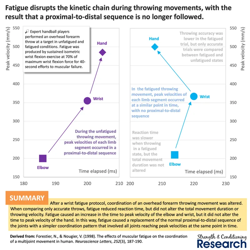
When we experience fatigue (through any mechanism), there is also a concurrent interference in our ability to coordinate movements. This then leads to a reduction in efficiency and hence a reduction in exercise performance. This interference in our ability to coordinate movements can be so severe that even fundamental movement patterns (such as proximal-to-distal sequences) are disrupted. Importantly, this fatigue mechanism does not itself cause any changes in muscle force production. It only reduces the efficiency of the movement pattern being performed.
.
#2. Reductions in central motor command generated (and hence in the ability to recruit high-threshold motor units)
During exercise, we tend to experience a reduction in the ability to produce a central motor command in the motor cortex, which we can call "supraspinal central nervous system (CNS) fatigue". Contrary to popular belief (both in exercise science and also in the wider fitness industry), this supraspinal CNS fatigue is not caused by producing a high level of central motor command, as can be seen by the fact that low-intensity exercise tends to be most effective at causing this type of fatigue mechanism to occur.
.
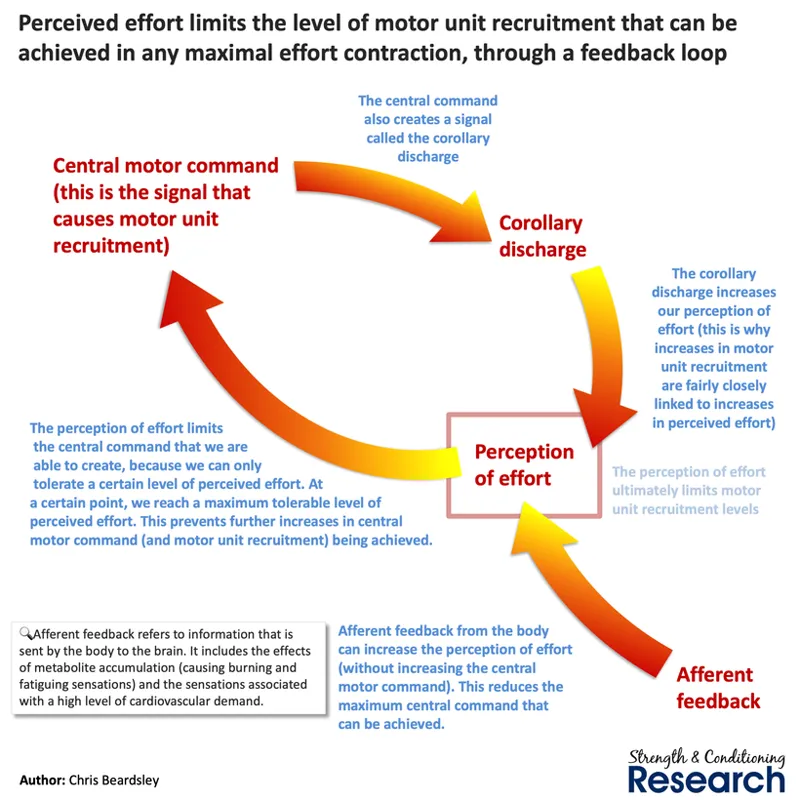
In fact, supraspinal CNS fatigue occurs because of feedback received from the body. This happens for three related reasons: [1] because there is a maximum tolerable perception of effort that we can withstand, [2] because the central motor command is limited insofar as it produces a copy of itself that is registered as a perception of effort (this is called the "corollary discharge), and [3] because other things can also contribute to the perception of effort, which then leaves less available capacity for the central motor command. Importantly, this fatigue mechanism reduces the level of high-threshold motor unit recruitment, preventing some of the most adaptable, fast twitch muscle fibers from being activated (and therefore trained).
.
Although many popular commentators in the fitness industry still either deny that CNS fatigue exists or claim that it is poorly understood, supraspinal CNS fatigue during exercise has actually been extremely well-described by the psychobiological model of fatigue that explains the interactions between perceived effort, motivation, and the central motor command.
.
#3. Reductions in central motor command transmitted (and hence in the ability to recruit high-threshold motor units)
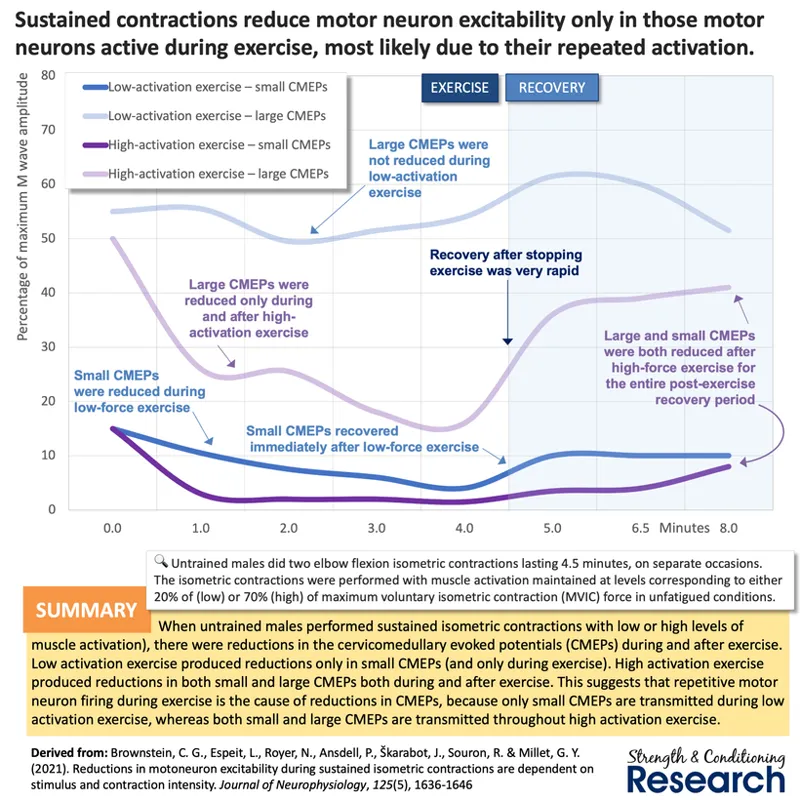
During exercise, we also tend to experience a reduction in the ability to transmit the central motor command signal down the motor neurons of the spinal cord. This appears to be a simple desensitization process in which the magnitude of the signal transmitted gradually reduces over the bout of exercise. This fatigue mechanism also reduces the level of high-threshold motor unit recruitment, preventing some of the most adaptable, fast twitch muscle fibers from being activated (and therefore trained).
.
PERIPHERAL FATIGUE MECHANISMS
#1. Reductions in cell membrane excitability
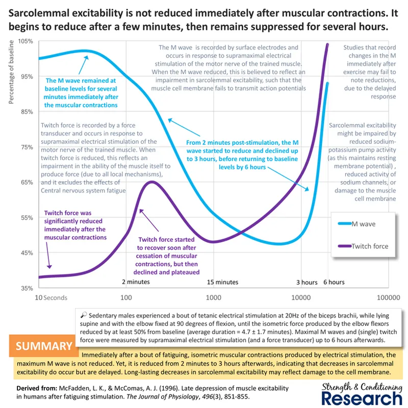
After the central motor command reaches the neuromuscular junction and activates the relevant muscle fibers belonging to the motor units that have been recruited, action potentials must travel along the cell membranes of the activated muscle fibers. For the muscle fibers to produce force, the cell membranes must transmit these action potentials to the transverse tubules and then onwards to the triadic junction so that excitation-contraction coupling can happen. Nevertheless, there is a fatigue mechanism that causes reduction in the ability of the cell membrane to transmit these action potentials. Since it is a slightly delayed mechanism, it is likely produced by a biochemical damage effect that takes time to occur.
.
#2. Excitation-contraction coupling failure (ECCF)
After the cell membranes have transmitted their action potentials to the transverse tubules, the signal arrives at the voltage sensor of the triadic junction, where excitation-contraction coupling can occur. Excitation-contraction coupling occurs when the voltage sensor interacts with the calcium ion store of the triadic junction, causing calcium ions to flow into the cytoplasm in response to the presence of an action potential being detected by the voltage sensor. Nevertheless, ECCF causes a reduction in the ability of the voltage sensor to interact with the calcium ion store, which occurs when calcium ions accumulate in the cytoplasm and stimulate the production of proteases.
.
#3. Loss of calcium ion sensitivity
After excitation-contraction coupling causes calcium ions to flow into the cytoplasm, the actin myofilament must detect them and then change its conformation to attract myosin. Nevertheless, there is a fatigue mechanism that causes reduction in the ability of actin to detect calcium ions. This seems to be a simple desensitization mechanism that occurs when too many calcium ions accumulate in the cytoplasm.
.
#4. Phosphate accumulation
After actin detects the presence of calcium ions, it attracts myosin to bind with it and produce a crossbridge. This crossbridge is what then generates mechanical tension and permits movement to occur. Since the crossbridge needs ATP to function, stopping ATP from converting into ADP and a phosphate can easily cause fatigue at this point. By accumulating inside the cytoplasm, phosphates can reduce the conversion of ATP into ADP and a phosphate since this chemical reaction is reversible.
.
#5. Acidosis
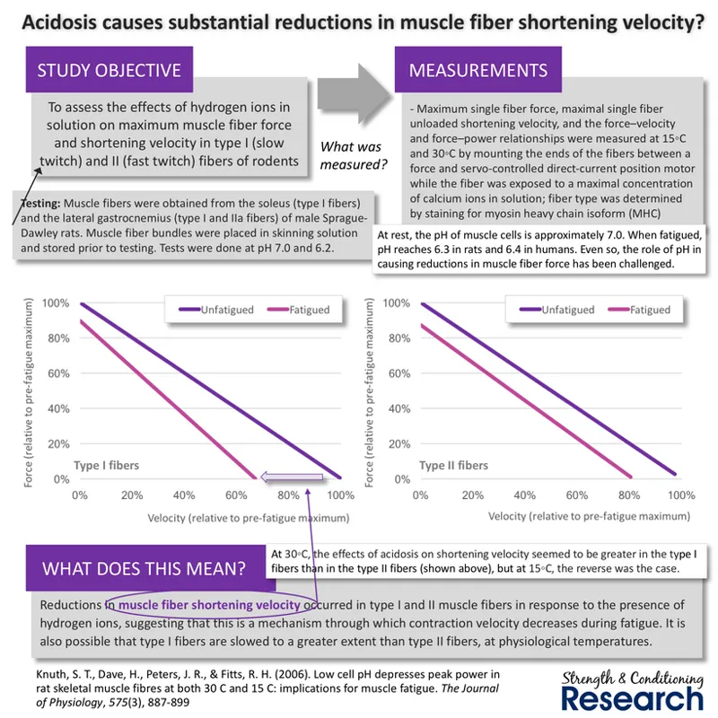
After a crossbridge has formed, it must detach so that the same myosin head can form another crossbridge further along the actin myofilament. However, acidosis can delay the detachment of the myosin head from the actin myofilament, which slows down the muscle fiber shortening velocity, but interestingly without affecting muscle fiber force very much at all.
.
Conclusions
Fatigue is a temporary and reversible reduction in exercise performance (often measured as a reduction in maximum strength) typically produced as a result of a previous but of exercise. Fatigue can be observed during exercise, and in this case is produced by a number of different mechanisms. Inside the central nervous system (CNS), peri-workout fatigue can occur as a result of [1] alterations in the coordination pattern that is implemented, [2] reductions in the level of central motor command that is produced inside the motor cortex (called “supraspinal CNS fatigue”), and [3] reductions in the size of the central motor command that is transmitted down the motor neurons of the spinal cord (called “spinal CNS fatigue”). In the muscle itself, peri-workout fatigue can occur as a result of either calcium ion-related mechanisms or metabolite-related mechanisms. Calcium ion-related fatigue mechanisms can occur due to [1] damage to the muscle cell membrane causing reductions in the transmission of action potentials to the triadic junction, [2] damage to the minor proteins of the triadic junction causing excitation-contraction coupling failure, and [3] a reduction in the sensitivity of actin myofilaments to the presence of calcium ions. Metabolite-related mechanisms can occur either due to [1] phosphate accumulation reducing the conversion of ATP into ADP and a phosphate, or [2] the accumulation of hydrogen ions (acidosis).
Comments (4)
Hi Chris! I like very much this post. Why is linked ion related fatigue with low frequency? I don’t see the relationship. Thanks!!
Low-frequency fatigue is just the technical term for a measurement method that identifies the presence of calcium ion-related fatigue. It refers to the ratio of low-frequency stimulated force to high-frequency stimulated force.
Hi chris, i got a question regarding some informations that i often see online or during differents seminars about the relation between cortisol, fatigue and hypertrophy. It is often stated that too much volume, or too much intensivness (sets closer to faillure or heavy loads) increased the amount of cortisol during a workout, which make sense IMO since we need to mobilize energy in order to realize the given task (in this case, a strenght workout). Where im starting to get lost a bit, is that the « experts » mention that cortisol will negatively impact Mtor signaling by the activation of AMPK signaling which will impede the muscle growth. My first question is : 1) Is the production of cortisol during a workout can have that much of an impact on muscle hypertrophy ? Or is it only a physiological marker of an intense workout ? 2) if too much cortisol is indeed impeding muscle growth, could it also affect the recovery rate of a workout by delaying the stage of muscle repair and hence, creating a certain amount of CNS fatigue ? I know that calcium ion related fatigue is playing a big part in the latter, but i’m just trying to disentangle the truth from the false. Thank you for your time
Historically, the post-workout hormone elevations (testosterone and cortisol) were thought to be important mediators of the hypertrophy and atrophy processes, which is why some people still talk about them in that way. Even so, more recent research has shown that such hormone elevations are not important for muscle adaptations and probably serve other functions in the body instead. In practice, I don't consider the cortisol effects of workouts at all.